Skin Cancer Checks Sunshine Coast
Sunshine Coast Skin Cancer Check
Protecting your skin is vital, especially in sunny Queensland. At Coastal Skin & Laser on the Sunshine Coast, we offer skin cancer checks tailored to your individual needs.
Our services include:
- Full Body Skin Cancer Checks
- Mole Mapping with FotoFinder Technology
- Monitoring of Mole Changes with AI Assistance
- Preventative Skin Health Advice
- Skin Cancer Treatment Options (if required)
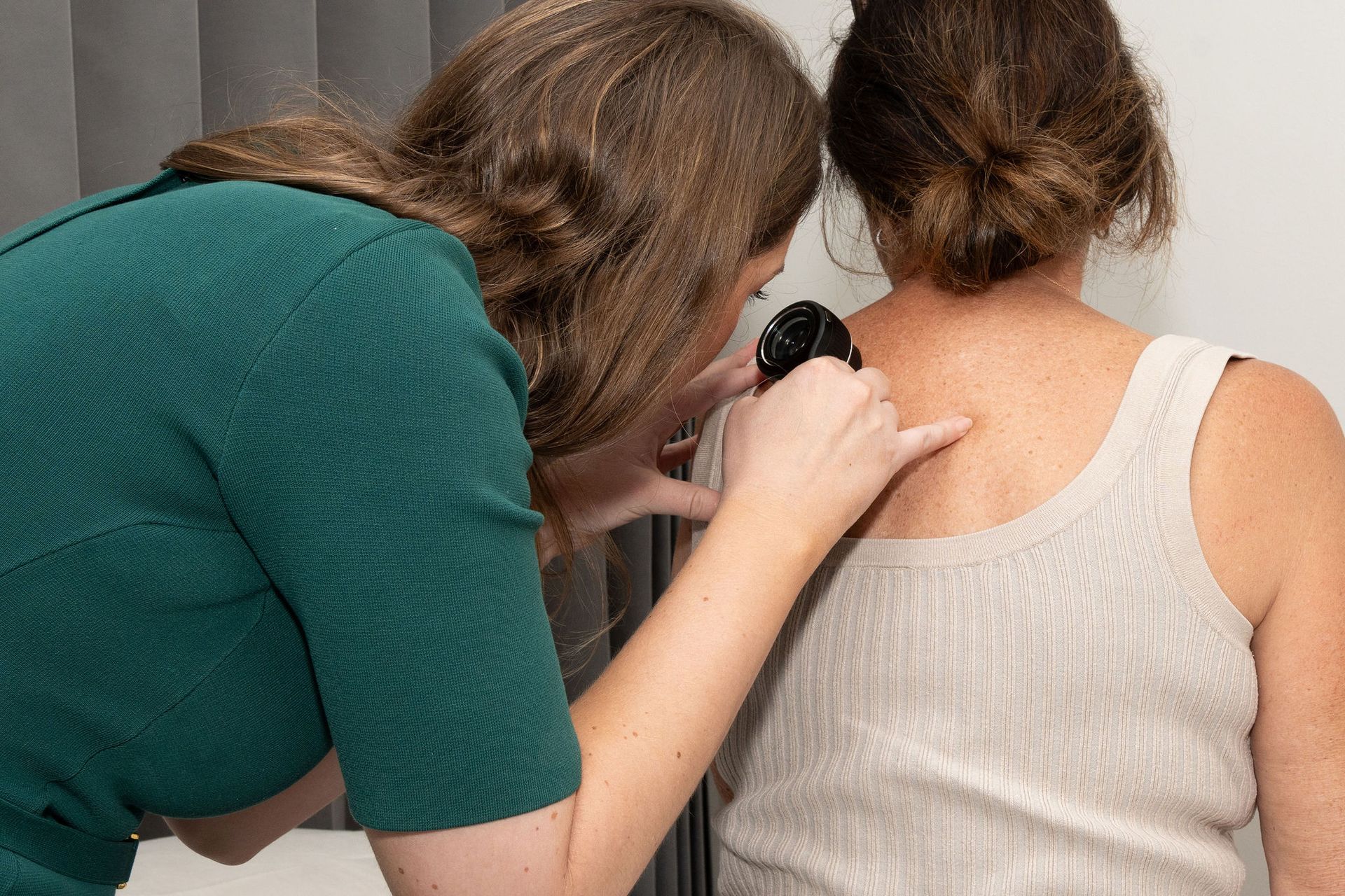
Each skin cancer check is conducted by a trained GP Specialist and Registered Nurse using advanced imaging tools to assess your skin from head to toe. With a focus on early detection, we support long-term skin health through professional assessment and ongoing monitoring.
As a skin clinic on the Sunshine Coast, we welcome patients from Noosa, Coolum, Maroochydore, Mooloolaba, Caloundra and surrounding areas.
To book a skin cancer check or speak with our team, call
(07) 5355 6033.
Total Body Mapping
Coastal Skin & Laser offers skin cancer detection using imaging and mapping tools, aiming to identify subtle changes over time. All assessments are led by GP Specialists with training in skin cancer detection, supported by mole mapping technology for comparisons and record keeping.
Our clinic uses FotoFinder technology to perform total body mapping and assist with monitoring skin changes over time. This supports clinical decisions around follow-up, removal or referral when required.
Each consultation includes a discussion, full skin check and digital records that can support long-term monitoring. If treatment is needed, we offer in-clinic options or referral to an appropriate specialist. By aiming for accuracy and continuity of care, we help support early detection and aim to reduce the risk of missed changes.
Book your skin cancer check with Coastal Skin & Laser through our online booking system today.
This is paragraph text. Click it or hit the Manage Text button to change the font, color, size, format, and more. To set up site-wide paragraph and title styles, go to Site Theme.
Price Guide
Note: If an excision or punch biopsy is required, an out-of-pocket fee will apply. Your doctor or nurse will advise you of this at the time.
Automated Total Body Mapping
ATBM (Automated Total Body Mapping) $220
Medicare card holders receive a rebate of $43.90.
Cryotherapy
Cyotherapy $75
This means the use of liquid nitrogen to treat pre-malignant lesions such as solar keratoses. Medicare cardholders will receive a rebate of $39.20. when 10 or more lesions are treated
Dermoscopy
Dermoscopy $95
This is a follow up appointment that may be required in between your annual body maps to track changes in pre-existing moles. Medicare card holders will receive a rebate of $43.90 for this service if a fee applies.
FAQs
What is body mapping?
Earlier detection of skin cancer with Automated Total Body Mapping
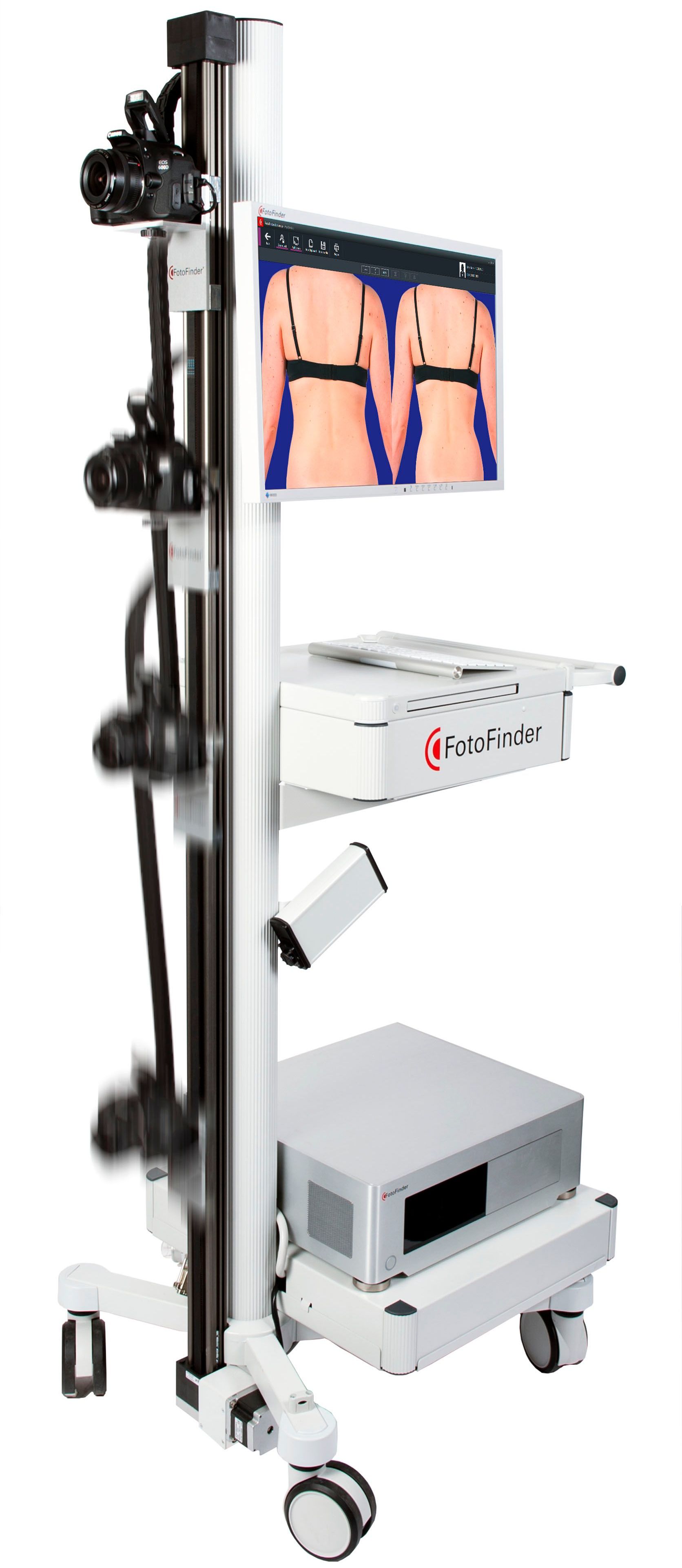
Skin cancer can be best managed if it is detected early enough. The biggest risk factors are excessive exposure to sunlight and genetic predisposition. For this purpose, we use the modern FotoFinder ATBM technology for Automated Total Body Mapping.
Did you know?
One Australian is diagnosed with melanoma every 30 minutes
For optimal safety, it is encouraged to track the entire skin in addition to individual moles. The advantages of the computer-assisted ATBM procedure: Allows entire skin documentation during regular check-ups. Every visit, new photos can be compared to past sessions allowing the visualisation of every change at the earliest possible stage.
A “map” of your skin: Automated Total Body Mapping
Imagine your skin as a landscape. Automated Total Body Mapping (ATBM) captures the entire skin surface in the long-term. We use the high-tech FotoFinder system to create a “body map” of your moles and take photographs of your body systematically—from head to toe and all sides—within a few minutes. We can obtain a full evaluation of new or changed lesions instantly during your checkup with the powerful FotoFinder Artificial Intelligence. With this software, you can be assured your data will be safe, secure and confidential.
Early diagnosis with technology: Video dermoscopy
In addition, suspicious moles are examined dermoscopically. With a special video dermatoscope, we take enlarged pictures of your moles which clearly show the pattern and structures. The digital storage of your skin images enables an objective comparison of previous and current skin findings during regular check-ups. You can follow your past and present examinations effortlessly on screen.
Keep the overview: Skin cancer prevention with FotoFinder
- Long-term and complete observation of the entire skin and every single mole
- Regular check-ups show new and changed moles at an early stage
- Mole-visualization from head to toe and analysis for malignancy
- Less unnecessary excision
- powered by FotoFinder Artificial Intelligence
Am I a high-risk patient?
If you can answer “YES” to any of these questions, please contact us!
- Do you have fair skin that is sensitive to sunlight?
- Do you have a particularly large number of moles?
- Do you have large congenital nevi?
- Do you have atypical moles or moles that have recently changed?
- Did you get sunburnt as a child or adolescent?
- Is there a history of skin cancer in your family?
- Do you have a previous history of having been diagnosed with skin cancer?
- Are you exposed to strong sunlight or UV rays at regular intervals?
Particular caution with:
- Newly developed moles
- Changes in colour, e.g. lighter, darker, new shades
- Increase or decrease of size, thickness or elevation
- Bleeding moles
- Changes in the surrounding area of moles, e.g. redness, white colouration, swelling
- Discomfort, e.g. itching, burning, foreign body sensation
How does it work?
Total body mapping is performed in our body mapping room by capturing images of all body areas. The camera slides up and down a stand for fast and consistent photography. Captured images are displayed immediately on-screen and directly and securely stored in the patient file.
When you return for your follow up images, comparisons are made and the technology will automatically highlight changes to pre-existing moles as well as detecting new moles.
After the mapping is complete, the doctor will assess the highlighted moles and use the powerful dermatoscope to capture up close images of any moles that look suspicious in nature.
A plan will then be made with you and your treating doctor on the course of treatment.
This can incorporate the following:
- Follow up dermoscopy (to monitor moles that don’t need excising, but need tracking)
- Biopsy of a mole (taking a sample and sending it to the lab for diagnosis)
- Excision (taking the whole lesion in our treatment room)
- Cryotherapy (using liquid nitrogen to ablate pre-malignant moles)
How do I prepare for my appointment?
- Remove make up and nail polish prior to your appointment.
- Avoid having a spray tan close to your appointment date.
- Avoid having IPL/Laser within one month of your appointment date.
- Ensure you have clean skin with minimal or no moisturiser.
- Wear light coloured under garments (briefs not boxer shorts for men).
- If you are hairy in areas which you have moles, you may consider waxing/shaving those areas 7 days prior to appointment time (to allow any skin reactions to heal).
- If you have any moles/spots you are concerned about, either mark them or photo them to remind yourself to ask the doctor.
- If you have had prior skin cancer excisions, it’s helpful to bring a copy of the pathology result with you.